top of page


Journal Covers
April 2011, Volume 10, Issue 4

The Beauty of Proteomics. 33" by 44", oil on canvas. Full description here.
On the cover of Cell, December 2007, 128: 59.

Dance of the Clock Gene Proteins. Oil on Canvas, 36" x 42". Full description here.
Cover of Preclinica, July/August 2004, Volume 2, Number 4.

Figures in a Beta Sheet. Oil and mixed media on canvas, 18" x 24", 2003. Full description here.
On the cover of Molecular and Cellular Proteomics, December 2012.

Proteasome Bashe Dragons Devouring and Digesting Ubiquinated Substrates. Digital collage of watercolor elements. Full description here.
Cover of the Fourth International Conference on Ubiquitin, Ubiquitin-Like Proteins, and Cancer. February 2008.

HUMO and SENP1 wrestling over the Goddess. Oil on canvas, 32" x 46". Full description here.
SUMO, Ubiquitin, UBL Conference Proceedings 2010

Digital collage of watercolor elements. Full description here.
On the cover of Molecular and Cellular Proteomics, Mach 2016, Volume 15, Issue 3, p753-1175

An Epigenetic Map 20” x 20” Oil and mixed media painting on board. Full description here.
Cover of Molecular and Cellular Proteomics, August 09, 2019,
Volume 18, Issue 8,

Through the Multiomix Looking Glass. Playing pieces modeled after characters in Lewis Carroll’s “Through the Looking Glass”, in a world that resembles a chess game, represent the many professional areas working towards the same goal; that of making sense of all the information they are uncovering in order to capture the knowledge needed to learn the answers to the questions humankind seeks.
On the Cover of American Scientist, May-June 2009.

The First World was Red. Dine' creation narrative and the evolution of the cell. See full description here.
Cover of Current Opinions in Structural Biology, 2000. Oil on Canvas, 33” x 44”, 2000

On the cover of Cell Metabolism, Feb. 25, 2013, Vol. 17, No.2.

Circadian Candy Clock. Watercolor and digital collage. The circadian clock plays an important regulatory role in a wide range of metabolic functions. The cover image illustrates how clock speed is regulated by glucose levels (candies) sensed via O-GlcNAcylation, as shown by Li and colleagues.
Cover of Cell, June 2008, volume 2, number 6.

Cells Both Dead and Alive as Calabash Fruit in the Tree Where Blood Moon is Impregnated by One Hunahpu, Oil on canvas, 33" x 44", 2000. Full description.
Cover of Nature Reviews Genetics, July, 2003.

Dawn of the Double Helix. Oil on canvas with gold leaf. Full description here.
Cover of the Monell Spring 2005 Colloquium.

Olfactory Garden One. Oil and mixed media on canvas, 18 x 24, 2004. Full description here.
Cover of Nature Reviews Genetics, May, 2001, Volume 2, Number 5.

Nanotechnology II. Oil and mixed media on canvas. See full description here.
Cover of Nature Reviews Genetics, July, 2001, Volume 2, Number 7

Nanotechnology I. Oil and mixed media on canvas. See full description here.
Cover of Nature Reviews Genetics, November, 2003.

Shiva as Telomerase inside of a Telomere Loop. Oil and mixed media on canvas, 2003, 32" x 38". Full description here.
Cover of Nature Reviews Genetics, June 2001, Volume 2, Number 6.

Nanotechnology III. Oil and mixed media on canvas. See full description here.
January 2002
Nature Reviews Molecular Cell Biology

Cell Signals and Mayan Legends, mixed media, 26" x 33", 2001. Description here.
Cover of Nature Reviews Molecular Cell Biology, December 2001, Volume 2, number 12.

FAS Sisters Receiving a Death Signal. Oil on board, 25" x 33", 2000. Full description here.
On the cover of Molecular and Cellular Proteomics, 2013, Volume 12, Issue 4, p833-1045

Glycomics Plate. Painted ceramic plate. Full description here.
On the cover of Molecular and Cellular Proteomics, June 2017, Volume 16, Issue 6, p959-1172

Proteomics Symphony. Oil on canvas, 24" x 36". Proteogenomics as an orchestrated analysis of DNA, RNA, protein and phosphoproteins.
Poster and program cover for the Tenth International Symposium on Mass Spectrometry in the Health and Life Sciences: Molecular and Cellular Proteomics, August 21-25, 2011.

The Tritons of Mass Spectrometry.2011. Oil on canvas, 24" x 30". See full description here.
On the cover of Molecular and Cellular Proteomics, March 15, 2019
Volume 18Supplement 1S1-S190

Tales of Four Conceptions. mixed media and digital collage.. Full description here.
On the cover of Molecular and Cellular Proteomics, December 2012.

Proteasome Bashe Dragons Devouring and Digesting Ubiquinated Substrates. Digital collage of watercolor elements. Full description here.
On the cover of Molecular and Cellular Proteomics, May 2021.

Glycoproteins Puzzle. Mixed media. Full description here.
On the cover of Cell July 3, 2013, Vol. 154. No. 1 pp. 1-252.

Interspecies Tempest. Watercolor and digital collage. Full description here.
Cover of Folha De S. Paulo, September 2004

Heading 4"The Transportation of Gilgamesh through an Ammonia Channel Into the Garden of the Gods", mixed media, 24 x 30, 2004. Full description here.
December 2006 Cover of
"Hommage Aux Origines de la Vie" by Chistine Dumitriu van Saanen

Spindles of Necessity V - Telophase. 3' x 4', oil on canvas, 2005. Full description here.
June 2004 Cover of Art Business News

Nanotechnology I. 20" x 24", mixed media, 1992. Full description here.
March 2010 29th Annual Gender Studies Symposium Lewis and Clark College

Dawn of the Double Helix. Oil on canvas with gold leaf. Full description here.
December 2003 Public Library of Science

Aquaporin. Digital collage of painted images and computer rendering of aquaporin protein. Aquaporin is a membrane protein that lets water into and out of the cell in a tightly controlled manner.
Cover of Thesis of Ruben A.T. Mars

RNA World. Oil and mixed media on canvas, 24" x 33", 2003, Full description here.
Cover of Preclinica, November/December 2003, Volume1, Number 5.

RNA World. Oil and mixed media on canvas, 24" x 33", 2003, Full description here.
Cover of "Structure" Structure, Vol 12, 1199-1207, July 2004.

Silenus Contemplating a Lipid Droplet Protein. Cover of "Structure" Journal, July 2004. Oil on canvas and digital collage. The obese Greek god Silenus contemplating the crystal structure of the lipid droplet protein TIP47. TIP47 contains a hydrophobic pocket that is conserved in the adipocyte lipid droplet protein, perilipin. Perilipin and its hydrophobic pocket are potential targets for anti-obesity drug design.
Structure of a Lipid Droplet Protein: The PAT Family Member TIP47
Sabrina J. Hickenbottom 1, Alan R. Kimmel 1, ConstantineLondos 1, and James H. Hurley. Structure, Vol 12, 1199-1207, July 2004.
Cover of The EMBO Journal, March 2001, Volume 20, Number 5. 4

The Transportation of Ra Through a Vault Protein out of the Night Sky and into the Birth of the Sunrise, 33" x 44", 2001,Tempera on Board. Full description here.
Tempest
More Information
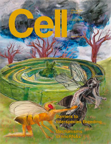
Interspecies Tempest description:
Developmental mechanisms generate interspecies hybrids that cannot propagate, as alluded to by the artist's depiction of the mythical Minotaur's labyrinth on the cover. However, even socially naïve animals rarely mate with members of other species. The mechanisms that control such behavioral reproductive isolation, an important tenet of evolution, are not well understood. As depicted on the cover, male flies tap other flies with their foreleg prior to attempting to mate. Fan et al. (pp. 89–102) show that foreleg removal permits males to mate with flies of other species. Activation of foreleg neurons expressing the chemoreceptor Gr32a is necessary and sufficient to inhibit interspecies courtship by D. melanogastermales. Gr32a is required to detect aversive hydrocarbons found on the cuticle of other drosophilid species, and one such hydrocarbon, z-11-pentacosene, is visible on the female fly on the cover. Strikingly, female flies utilize a Gr32a-independent mechanism to reject males of other species, with the cover depicting the female fly rejecting the male who is tapping her.
Dragon
Description of Ubiquitin Dragon:
In the cytosol (the ocean region in the painting) certain protein “fish” have been tagged for degradation by a seaweed-like chain of ubiquitin wrapped around their tails. One such fish is being shuttled towards the proteasome “dragon” by a fish-tailed sea god, while others are drifting towards destruction on their own. After digestion, the protein “fish” can be seen expelled from the other end of the dragon as bones. In Chinese mythology, Bashe Dragons are said to eat elephants and excrete their bones years later. In the nucleus, the bright orange land of the underworld in the painting, the black silhouette of a second proteosome dragon can been seen involved in the same task. This dragon is similar, but not the same, as the one in the cytosol in ways that are not yet understood.

Glycoprot

Description of Glycoprotein Puzzle
This art highlights from top to bottom the three main topic areas covered by the Special Issue. The first topic area is capturing glycoproteins/glycopeptides (in the purple under shading at the top right) away from the non-glycosylated species (in the green under shading at top left). Then the purple arrow represents introduction into a mass spectrometer for analyses by fragmentation techniques (shown in the yellow under shading region in the middle of the art piece) which is the second major topic area covered in the review. Finally, the third topic area of post-acquisition data analyses (shown in the blue under shading region at the bottom) takes the data generated (yellow arrow) and reassembles the pieces to map sites of modification and determine site-specific microheterogeneity (represented by the moss like structures) on the protein backbone (amino acids displayed as stones across the bottom of the picture).
EpiMap

Description of An Epigenetic Map
This painting is part of a series in which the artist explores proteomics, and stems from a collaboration with MCP-editor-in-chief Al Burlingame and associate editor Ralph Bradshaw, with input from Benjamin A. Garcia, Ph.D., Presidential Associate Professor and Simone Sidoli, postdoctoral scholar of the University of Pennsylvania; and Michael Washburn, Director of Proteomics, of the Stowers Institute for Medical Research. Description: In this Epigenetic Map, a phrase derived from CH Waddington's concept of the Epigenetic Landscape, a chromosome-shaped island in the center has both inaccessible mountainous terrain and flat open areas. Running over the landscape is a red ribbon of DNA, wrapped around clusters of grapes that represent histones incorporated into nucleosomes, and decorated with leaves of various colors representing epigenetic markers. Around the outside of the island, the accessible DNA of the flat, open area of the map has been magnified, so we can see epigenetic modifications to proteins and DNA being performed by mythological characters representing the enzymes that carry out this process. Starting in the lower left, the red DNA strand emerges from a tight clump of inaccessible chromatin, buried in red and orange leaves which represent repressive and transient histone markers. Hermes, the Greek winged messenger god, is decorating the histones with green, activating marker leaves. In his hand, he holds a message to activate MLL, an example gene product and epigenetic modifier. The shields represent proteins, and are labelled with the gene product they represent. You can see the Set/MLL label on the blue shield next to Hermes. Above him, a centaur is painting the grapes purple, indicating fully active histones. The centaur represents the unknown control mechanism of the epigenetic process. Across the top, two DNA modification enzymes are represented by the winged female figures above the unfurled and enlarged portion of the red DNA strand, where DNA bases A, G, T, and C can be read. The more sinister, bird-bodied figure on the left is oxidizing methyl groups, which modifies expression of the gene, while the more pleasant character on the right is adding methyl groups to the cytosines on the DNA. Continuing clockwise, Aglaia, goddess of beauty standing on the compass, applies enzyme modifications to the various protein shields in the form of brightly colored jewels. Below her, the satyr applies transient histone markers represented by orange leaves, while his female muse applies repressive histone markers in the form of red leaves. At the bottom right, the DNA again dives into a tight clump of red and orange leaves, where it is no longer accessible, like the DNA in the mountains on the chromosome island in the center.
Tritones

Description of Tritons of Mass Spec.
Tritones are sea gods which normally do the bidding of Poseidon, king of the seas in Greek Mythology, but they have come to San Francisco for The 10th Annual Symposium on Mass Spectrometry. The merman is tossing up strands of jewels, which represent proteins, towards the surface of the bay. The mermaid holds a shell that allows a stream of water droplets through a hole in the center, alluding to the principal of mass spectrometry whereby a stream of water droplets containing the proteins of interest are sent into an electric field and become charged. Once the droplets containing protein "necklaces" reaches the sky, they are struck by a bolt of lightning by the helpful Zeus. This reflects the step in Mass Spectrometry where electricity is used to chemically break the protein into smaller, ionized pieces, which are sorted and analyzed according to their charge and weight. Strands of pearls attached to large gems represent epigenetic modifications to the proteins, a valuable piece of information gathered using this technique.
Conceptions
Description of Conceptions.
Pictured is a metaphorical depiction of the different mechanisms of fertilization in four different groups of organisms. At the top are insects, represented by Eros and Psyche. In Greek myth, Eros keeps Psyche in his home in a big mountain. He comes to her in the night through the mountain. Insect eggs have a pore through which only one sperm can fit. The sperm is so large, it physically keeps competitors from entering. In the right quadrant, mammals are represented by Penelope and Odysseus, with the many suitors that she keeps at bay until her husband’s return locked outside the doors. In mammals, only one sperm gets into the egg, after which the outside of the egg will no longer permit any more sperm to enter, thus “shutting the door” on any competitors. In the lower quadrant, avians are represented by Eos, the rosy-fingered goddess of the dawn who rises into the sky from the river Okeanos at the start of each day. She has an unquenchable desire for handsome young men. The avian egg allows many sperm to enter, but only one does the actual fertilizing. At some point, the egg surface changes, and no more sperm are allowed to enter. The river, thick with reeds, represents a barrier to the sperm that have not arrived in time, symbolized by the men on the other side of the river, who were not able to make it across before the chamber of Eos is full. On the left, echinoderms are represented by Danae, who has been locked below ground by her father, King Acrisius, to keep her from getting pregnant with a child that has been prophesied to kill him. Zeus manages to impregnate her by turning into a golden shower and penetrating the fortress. Echinoderms release many sperm into the water, but only one finds its way into an egg.

Glycomics

Description of Glycomics Plate.
A cell membrane constructed of the half human, half fish (both water loving and land creature) Oannes of Mesopotamian mythology divides the image into two worlds - that of the cytoplasm (black) below the outside the cell (red) above. In the cytoplasm reside the water creatures - species of fish and sea monster. They are glycosylated, if at all, with a single sugar GlcNAc, represented by the blue and white dragonfly (in the larvae stage in the water and as a dragonfly on the outside of the cell). Spanning the membrane are various characters from Greek mythology representing membrane proteins. They all hold or are attached to a straight or branched object, which represent straight or branched sugars. At the end of the chain of sugars is a purple bird, representing Neu5Ac. The objects in these glycans, like the bird, are colored to reflect the “Recommended symbols and conventions for drawing glycan structures” published in the Essentials of Glycobiology*. Hera (or Juno) holds her golden apple tree full of Gal, GalNac, and GalN. Cerberus sports a snake on each head, which might be a short chain of IdoA and GalA, all with the purple bird on the end of each branch (Neu5Ac), while the ram with the golden fleece has only a single GlcNAc, reflecting the possibility that proteins in the cytoplasm may be glycosylated in this way as shown recently in the literature. For details, see Essentials of Glycobiology, 2009, 2nd edition, Varki A, Cummings RD, J.D. Esko, et al., eds., Cold Spring Harbor Laboratory Press, Cold Spring Harbor, NY.
Hypoxia

Description of HUMO and SENP1 wrestling over the Goddess of Hypoxiaypoxia
HIF1alpha stands on a high mountain peak, where the air is thin (hypoxic), sending out newly formed blood vessels from her gown and blood cells from her sleeves. SUMO is attempting to grab the Goddess to throw her to the Dragon (proteasome), lurking below them in the clouds. SENP1 wields a sword trying to attack SUMO, separating him from the Goddess and saving her from the jaws of the dragon. This piece was a special commission by Dr. Edward T.H. Yeh. It is based on the paper (Cell article 131:584, 2007), "SUMO-specific protease 1 is essential for stabilization of HIF1alpha during hypoxia." Cheng J, Kang X, Zhang S, Yeh ET.
UbiquitinProc

Description of Ubiquitin Conference Proceedings:
Ubiquitin-mediated proteasomal degradation (Center): The dragon (proteasome) breaks up a stretch of polypeptide, tagged for degradation with a string of gold medallions (ubiquitin) bearing the image of Alfred Nobel to symbolize the award of the Nobel Prize in 2004 to Avram Hershko, Aaron Ciechanover, and Irwin Rose. The center card is surrounded by keys that open up locks in the surrounding cards.
Starting from upper right in clockwise rotation:
SUMO-mediated proteasomal degradation: Based on the oil painting SUMO and SENP1 wrestling over the Goddess of Hypoxia (HIF1a), 2007 The Goddess stands on a high mountain peak, where the air is thin (hypoxic), sending out newly formed blood vessels from her gown and blood cells from her sleeves. SUMO is attempting to grab the Goddess to throw her to the Dragon (proteasome), lurking below them in the clouds. SENP1 wields a sword trying to attack SUMO, separating him from the Goddess and saving her from the jaws of the dragon. Cell 131:584, 2007.
Immune response: The Inca rose from a waning tribe to an empire through "strong leadership, strategic alliances, and skill in warfare" (Gods of Order, Gods of War, A Comparison of the Major Gods and Myths of the Inca and Aztec, by Gini Graham Scott), in much the same way developing a good immune system boosted survival of organisms.
Neurological diseases: After the painting Nerve Eye on Channel Two, 40" x 40", 1992.
Cardiovascular diseases: Heart was rendered using watercolor.
Cell division: From Spindles of Necessity #3 (Anaphase) oil on canvas 3' x 4'.
Circadian rhythm: From “Dance of the Clock Gene Proteins”, . Cell 128: 59, 12 2007.
Signal transduction: From Cell Signals and Mayan Legends, 26" x 33", 2001. Growth hormone binds to the receptor tyrosine kinase (blue house structure) which attracts the protein grb2 (grandmother). This triggers a series of events which activate the proteins necessary for cell division.
DNA repair: Isis is an ancient Egyptian mother goddess associated, among other things, with healing.
DNA replication: Stringing beads is an ancient past-time. Egyptians were stringing nucleic acid and phosphate backbone beads during DNA replication.
Cancer: Rendering of cancer cell after image courtesy of Zena Werb, UCSF.
bottom of page