
Cells, More Cells, and Making More Cells

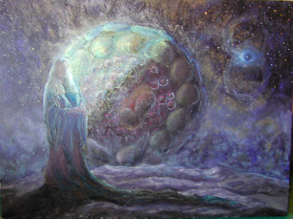
Plight of the Estrogen Ladies. 30" x 40", oil on canvas, 1991. Private collection.
The cycle of estrogen takes this small molecule through the bloodstream until it reaches cells such as uterine cells. An estrogen molecule has just transformed into the floating figure. She is attracted to the lotus flowers, which represent estrogen receptors on the surface of a cell. The flower captures her, and together they enter the cell and then the red cell nucleus. They then are bound to the purple DNA inside the nucleus. At this point, they cause the DNA to ultimately produce substances that cause the feelings and actions that estrogen is meant to trigger. Estrogen causes many, many things to occur.
Reference for description, "Biochemistry",Lubert Stryer, p. 1001, 1988.ISBN 0-7167-1843-X.

Ovariation. 30" x 40", oil on canvas, 1999. Private collection.
This painting was inspired by an image of a drosophila ovary releasing lots of eggs on its surface. From here, it became a human ovary. In humans, there is a large searching tentacle, called a fimbriae, which sweeps over an ovary searching for an ovulated egg. Once it finds the egg, it draws it into the fallopian tube. This egg has released an abnormal amount of eggs.

Conception. 30" x 40", oil on canvas, 1990. Private Collection.
This painting was inspired by an electron microscope image of a drosophila sperm fertilizing an egg. Microscope image taken and processed in the labs of Professor John Sedat and Professor David Agard, University of California at San Francisco
Woman Contemplating the Increasing Speed of her Biological Clock. 33" x 44", oil on canvas, 1999. Private collection.
At the time of this painting, it was thought that a woman is born with all of her eggs, in the thousands, (oocytes) at birth, and during her lifetime, she will ovulate only about 400 eggs. The rest of these eggs seemed to self-destruct via the mechanism of apoptosis, or programmed cell death. What causes these eggs to die? If they died at a constant rate until menopause, a woman would be losing two eggs a minute. It seems, however, that we lose fewer eggs during the early part of our life, and this egg loss gets faster and faster as a woman ages. The oocyte in the background is like a waning moon. Egg loss has reached the tenth hour of her biological clock.
Inspired in part by the paper "Oocyte Apoptosis: Like Sand through an Hourglass", Yutaka Morita and Jonathan Tilly, Developmental Biology 213, 1-17, 1999. Figure originally in "Prolongation of ovarian lifespan..." by Perez et. al, Nature Genetics. vol21 no. 2, 1999. Special thanks to Chris Gralapp.

The Beauty of Proteomics. 32" x 38", oil on canvas, 2011.
Angela Hvitved of Molecular and Cellular Proteomics writes:
It may be beautiful, but the April cover of Molecular and Cellular Proteomics is not just another pretty picture. The cover artwork, commissioned by MCP, is an oil painting by artist Julie Newdoll titled “The Worlds of Proteomics.” The painting is the first of a series in which the artist explores proteomics and stems from a collaboration with MCP co-editors Al Burlingame and Ralph Bradshaw. Newdoll worked with Burlingame and Bradshaw to learn about the many aspects of proteomics research and to understand the broad scope of science that MCP publishes. Their goal in initiating the series is to promote a deeper, richer appreciation for the field by those directly and indirectly involved with proteomics research.
Newdoll has a master’s degree in medical illustration and previously worked as a visualization specialist at the University of California, San Francisco. She found that fusing the powerful and beautiful concepts of science with cultural references both ancient and contemporary provided a new lens for creating her own view of the fascinating world of science. Other commissioned paintings by Newdoll can be found at her website.
The painting on the cover is accompanied by the artist’s description, which explains Newdoll’s inspiration for the piece:
“When proteins are made in the cell in response to some stimuli or event, they are targeted via an address system for a specific location or locations. In this painting, the various areas in a cell are represented by various worlds – there is the world of the sea in the cytosol, that of the air outside the cell and land or earth inside the nucleus. Inside the mitochondria and the endoplasmic reticulum, little islands have their own color scheme. A protein meant to be secreted to the outside of the cell follows an elaborate path of production, first forming inside the endoplasmic reticulum, later packaged into a membrane bubble which melds with the Golgi, and finally repackaged and released to the outside of the cell.
“From the artist’s perspective, a protein meant for the outside of the cell is rendered as a flying creature in this painting. It never ends up in the deep sea environment of the cytosol, or it would ‘drown.’ Likewise, proteins destined for the deep sea of the cytosol could not breathe outside the cell in the open air. And then there are the amphibians …”
“The Worlds of Proteomics” represents the broadest perspective of proteomics research, and Newdoll intends to use it as the foundation of the series. “The first in the series displays all realms of proteomics, while future paintings will zoom in on the various worlds and microcosms mapped out here,” she explains. The co-editors intend to feature future pieces on the journal’s cover to highlight specific areas of research.
To celebrate the debut of the new cover artwork, MCP, normally an online-only publication, has printed 500 copies of the April issue as well as a poster of the painting for distribution at the ASBMB annual meeting on April 9 – 13 and to members of its editorial board.

Sperm Surface Dress. 4' x 5', mixed media, 1994.
The patterns in the window and on the dress are from electron microscope images of the surface of a sperm.
Electron Microscope Images supplied by Dr. Dan Friend, Harvard Medical School, Pathology Department.

Spermbrella. 4' x 5', mixed media, 1994.
The sickle-like shape is taken from an electron microscope image of a guinea pig sperm.
Microscope image supplied by Dr. Dan Friend, Harvard Medical School, Pathology Department